
SEMIR: Semantic Minor-Induced Representation Learning on Graphs for Visual Segmentation
Abstract
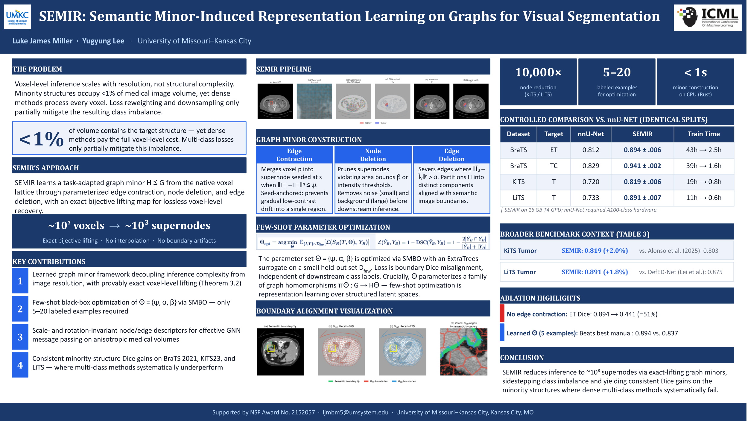
Segmenting small and sparse structures in large-scale images is fundamentally constrained by voxel-level, lattice-bound computation and extreme class imbalance--dense, full-resolution inference scales poorly and forces most pipelines to rely on fixed regionization or downsampling, coupling computational cost to image resolution and attenuating boundary evidence precisely where minority structures are most informative. We introduce \textbf{SEMIR} (\emph{Semantic Minor-Induced Representation Learning}), a representation framework that decouples inference from the native grid by learning a task-adapted, topology-preserving latent graph representation with exact decoding. \textbf{SEMIR} transforms the underlying grid graph into a compact, boundary-aligned graph minor through parameterized edge contraction, node deletion, and edge deletion, while preserving an exact lifting map from minor predictions to lattice labels. Minor construction is formalized as a few-shot structure learning problem that replaces hand-tuned preprocessing with a \emph{boundary-alignment objective}: minor parameters are learned by maximizing agreement between predicted boundary elements and target-specific semantic edges under a \emph{boundary Dice criterion}, and the induced minor is annotated with scale- and rotation-robust geometric and intensity descriptors and supports efficient region-level inference via message passing on a graph neural network (GNN) with relational edge features. We benchmark \textbf{SEMIR} on three tumor segmentation datasets—\textbf{BraTS 2021}, \textbf{KiTS23}, and \textbf{LiTS}—where targets exhibit high structural variability and distributional uncertainty. \textbf{SEMIR} yields consistent improvements in \emph{minority-structure Dice} at practical runtime. More broadly, SEMIR establishes a framework for learning task-adapted, topology-preserving latent representations with exact decoding for high-resolution structured visual data.
Lay Summary
Medical images such as CT and MRI scans can contain millions of voxels, but important structures such as tumors may occupy only a very small part of the image. This makes segmentation difficult because standard methods must process the full image grid and can miss small structures under severe class imbalance. SEMIR addresses this by converting the image grid into a smaller graph representation whose regions are aligned with meaningful boundaries. A graph neural network then makes predictions on this compact representation, and the results are mapped exactly back to the original image. Across brain, kidney, and liver tumor segmentation benchmarks, SEMIR improves segmentation of small tumor structures while reducing the amount of computation needed for prediction.