
S2M-Net: Spectral-Spatial Mixing with Morphology-Aware Adaptive Loss for Medical Image Segmentation
Abstract
Lay Summary
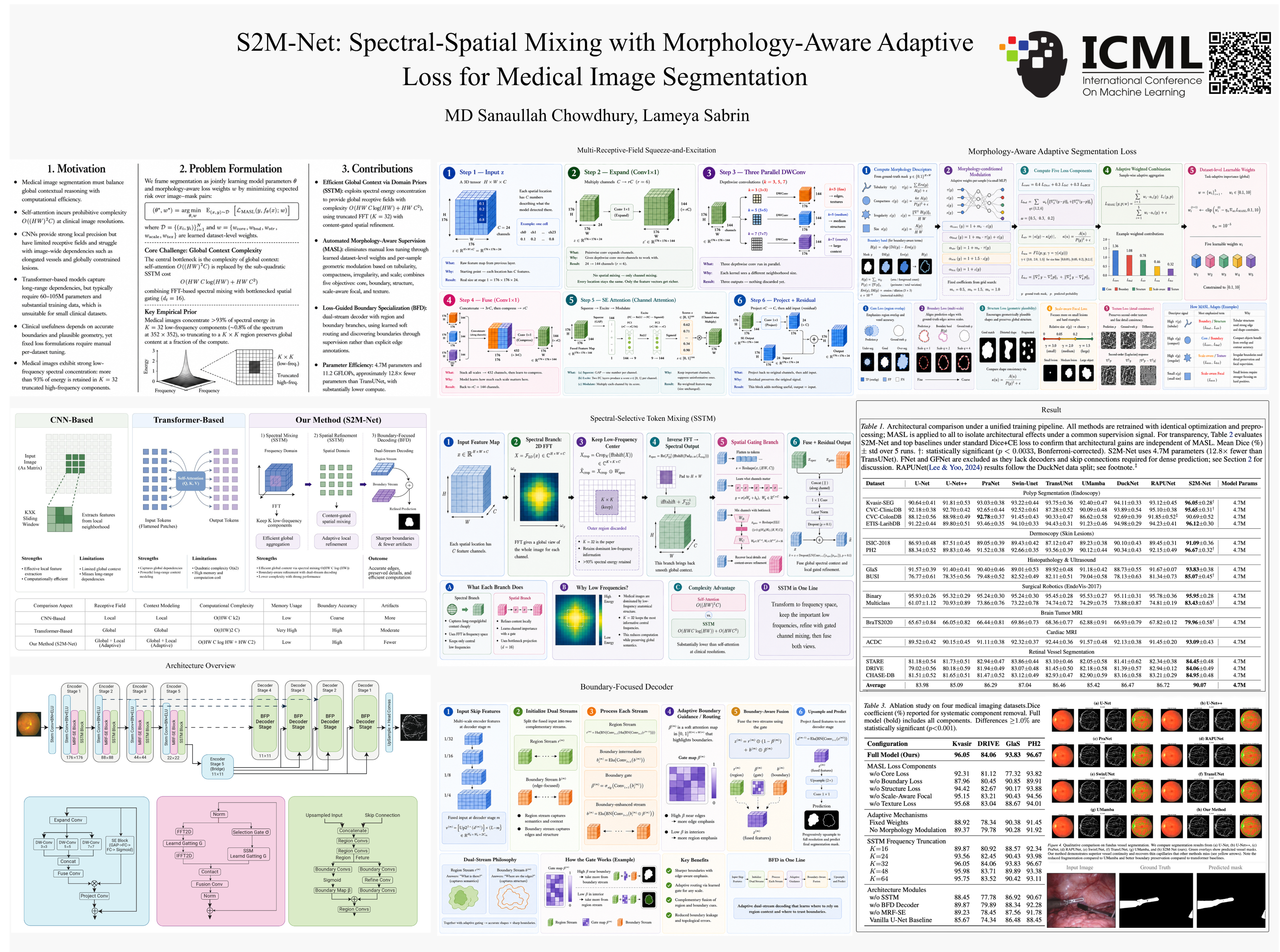
Medical image segmentation (the task of precisely outlining organs, tumors, or surgical instruments in scans) is a cornerstone of modern clinical care, supporting everything from cancer diagnosis to robotic surgery planning. Today's best AI models for this task borrow heavily from large language model technology, using mechanisms that examine every part of an image in relation to every other part simultaneously. This is powerful, but extraordinarily expensive: as image resolution grows, computational cost explodes, putting these tools out of reach for hospitals with limited hardware. We introduce S2M-Net, a compact AI model that achieves state-of-the-art segmentation accuracy while using up to 12.8 times fewer parameters than leading alternatives. Our key insight is that medical images (unlike natural photographs) concentrate most of their meaningful information in low frequency patterns, the broad shapes and smooth gradients that define organ boundaries. By working primarily in the frequency domain and processing only the most informative signal components, S2M-Net captures the global picture efficiently without the computational explosion of conventional approaches. Beyond architecture, we introduce a smart training objective that automatically adapts to the geometry of whatever structure is being segmented, behaving differently for thin blood vessels, irregular tumors, and compact polyps, without requiring manual tuning for each new dataset. Tested across 15 diverse medical imaging datasets spanning MRI, CT, ultrasound, endoscopy, and retinal imaging, S2M-Net outperforms all baselines on 14 of 15 benchmarks. Its small footprint makes it deployable on standard clinical workstations and edge devices, broadening access to AI assisted diagnostics in resource limited healthcare settings worldwide.